Emulate cell culture system ( organ on a chip)
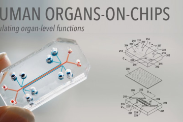
The Emulate cell culture system, also known as an "organ-on-a-chip" platform, is a cutting-edge microfluidic technology that replicates the physiological environment and mechanical forces experienced by cells in human organs. By incorporating microengineered chips lined with living human cells, the system enables researchers to study complex biological processes, disease mechanisms, and drug responses in a more predictive and human-relevant manner than traditional in vitro models. Emulate's organ-on-a-chip technology integrates key features such as dynamic fluid flow, tissue-tissue interfaces, and mechanical cues, making it an invaluable tool for biomedical research, drug development, and personalized medicine applications.